38 heart diagram with labels and blood flow
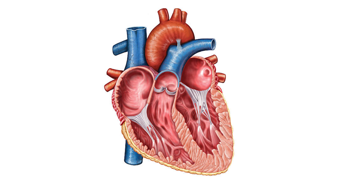
The Heart | Circulatory Anatomy - Visible Body One chamber on the left receives oxygen-rich blood from the lungs and another pumps that nutrient-rich blood into the body. Two valves control blood flow within the heart's chambers, and two valves control blood flow out of the heart. 1. The Heart Wall Is Composed of Three Layers. The muscular wall of the heart has three layers. How the Heart Works: Diagram, Anatomy, Blood Flow - MedicineNet The heart is an amazing organ. It starts beating about 22 days after conception and continuously pumps oxygenated red blood cells and nutrient-rich blood and other compounds like platelets throughout your body to sustain the life of your organs.; Its pumping power also pushes blood through organs like the lungs to remove waste products like CO2.; This fist-sized powerhouse beats (expands and ...
Anabolic steroid - Wikipedia In addition to morphological changes of the heart which may have a permanent adverse effect on cardiovascular efficiency. AAS have been shown to alter fasting blood sugar and glucose tolerance tests. AAS such as testosterone also increase the risk of cardiovascular disease or coronary artery disease.
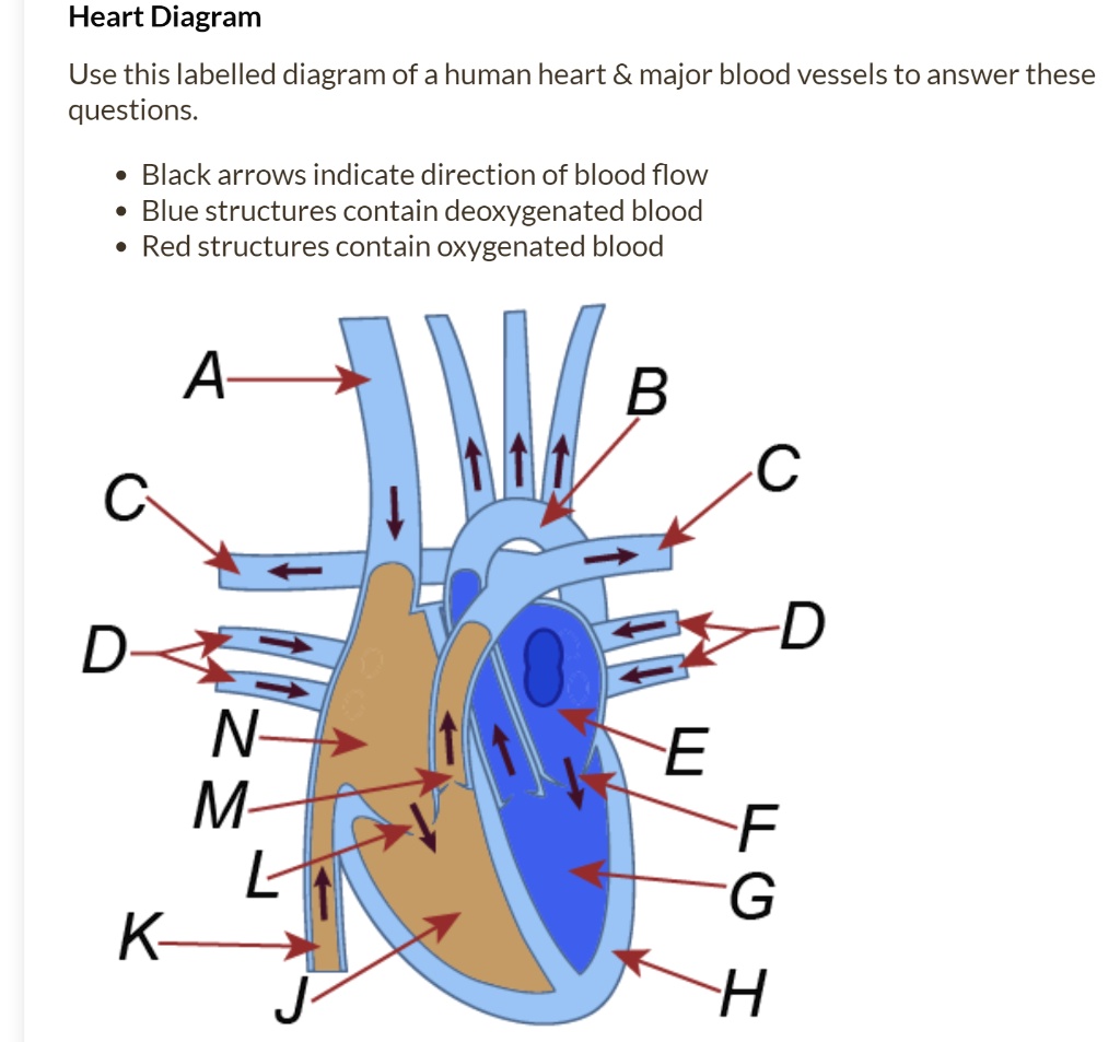
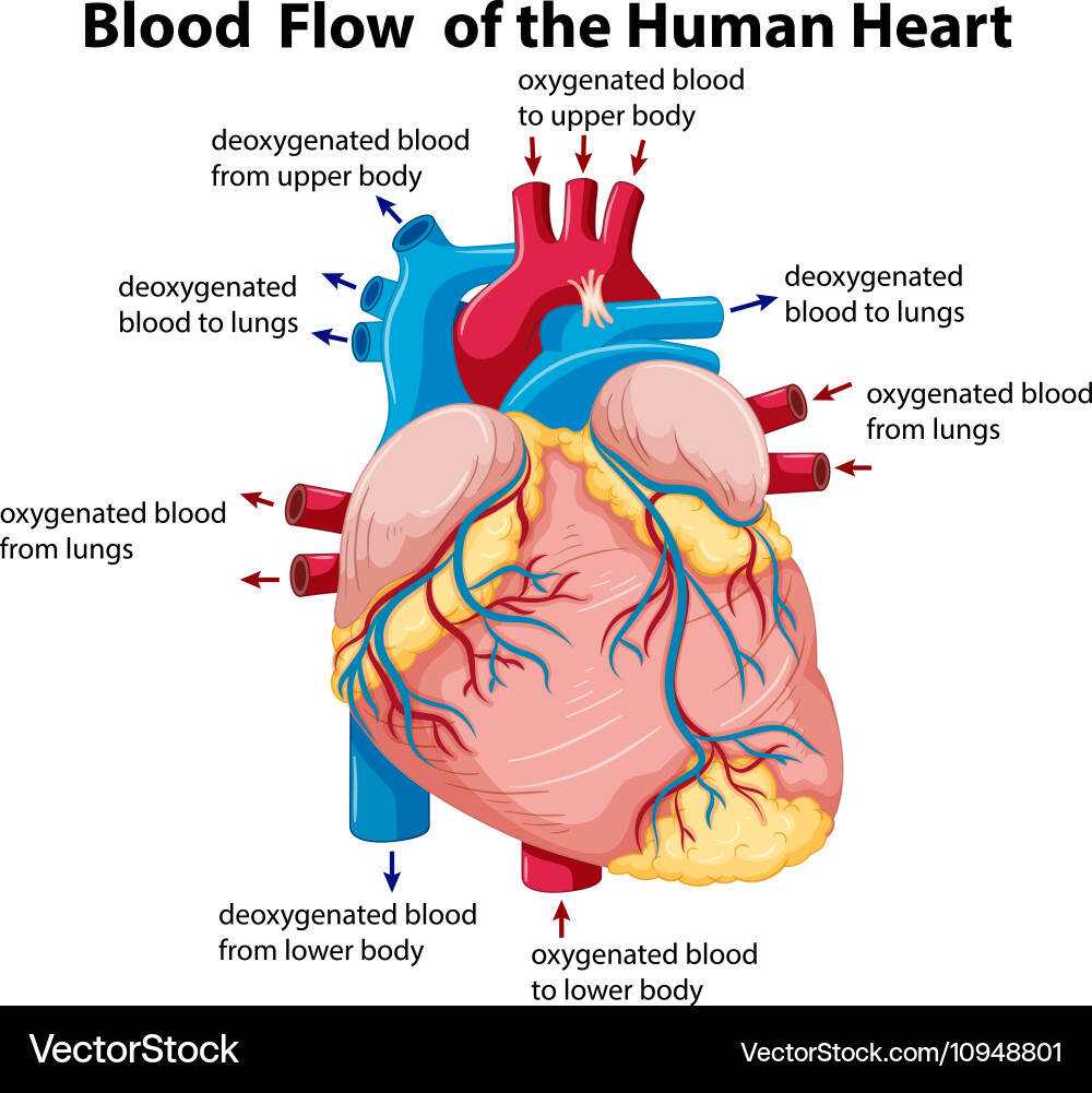
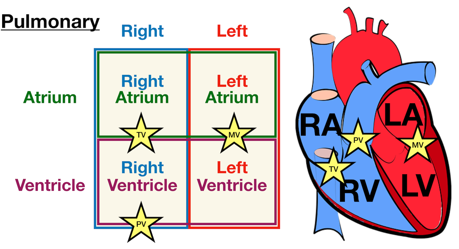
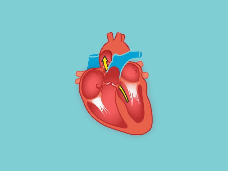
Heart diagram with labels and blood flow
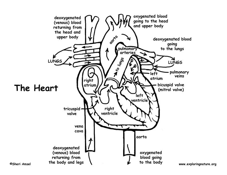
Human Heart Diagram Labeled | Science Trends Let's examine the anatomy of the heart along with some diagrams that show how the heart operates. Anatomy Of The Heart The human heart usually weighs somewhere between 10 to 12 ounces in men and between 8 to 10 ounces in women, and in terms of size is roughly the size of the fist. How the Heart Works How Blood Flows through the Heart Arteries take blood away from your heart. Your heart valves help control the direction the blood flows. Heart valves Heart valves control the flow of blood so that it moves in the right direction. The valves prevent blood from flowing backward. The heart has four valves. The tricuspid valve separates the right atrium and right ventricle. Heart Diagrams for Labeling and Coloring, With Reference Chart and Summary (Not for black and white photocopying). - One black and white heart diagram with lines for students to fill in labels, and arrows showing blood flow - One black and white heart diagram with no lines or labels, but arrows included, so you can customize what labels the diagram will include
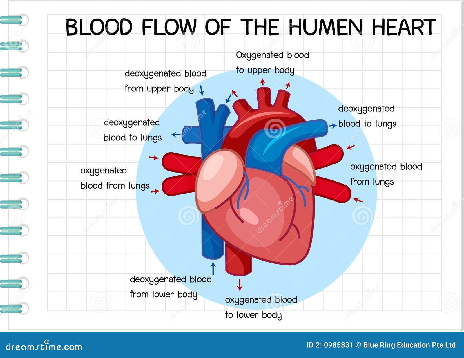
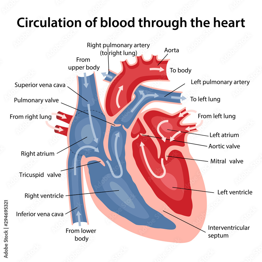
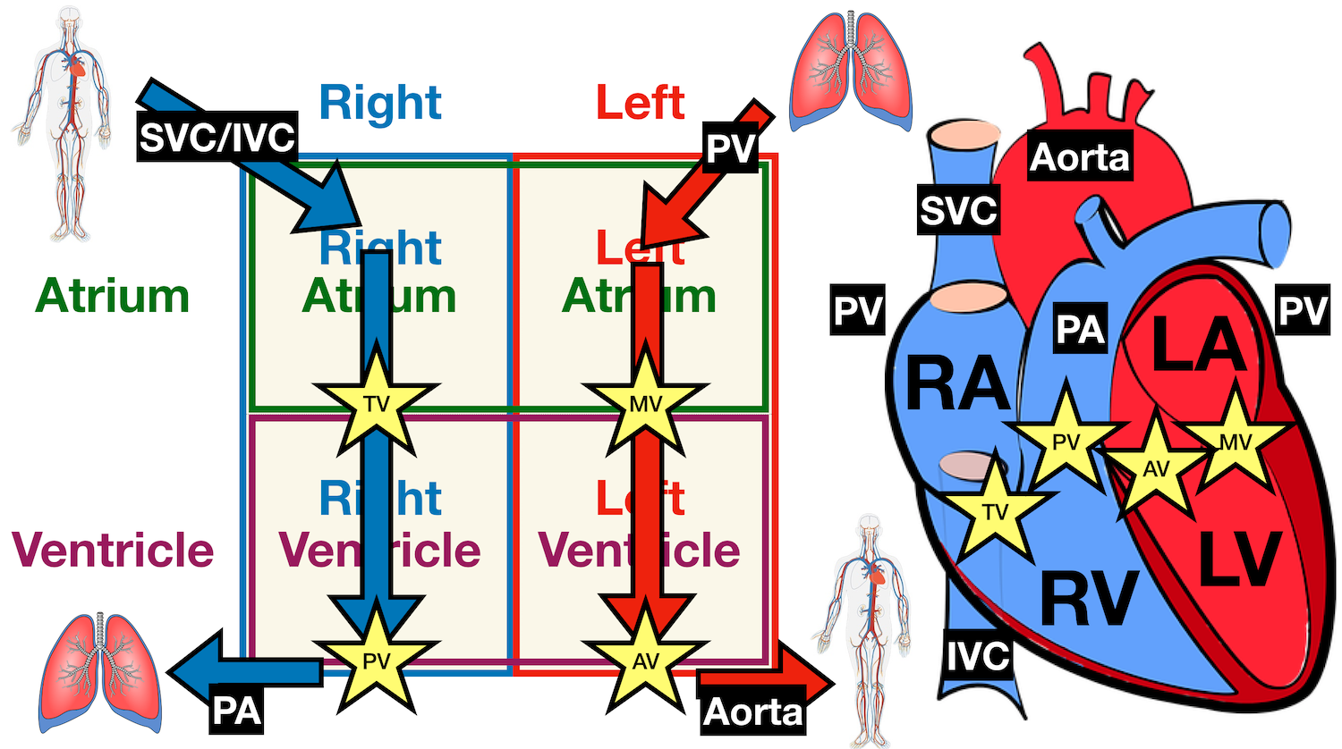
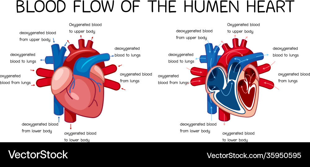
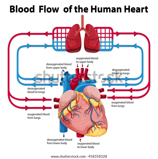
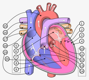
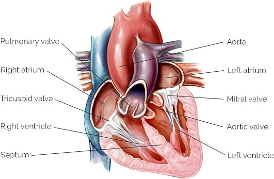
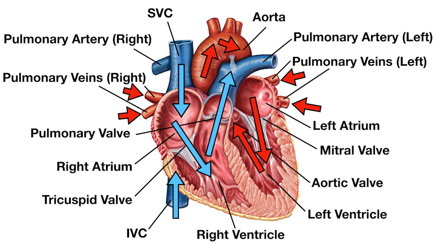
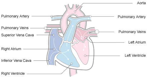
Heart diagram with labels and blood flow. Blood Flow Through the Heart: Pathways and Circulation - Cleveland Clinic Blood flows through your heart through a series of steps. These steps take place in the space of one heartbeat — just a second or two. On the right side Oxygen-poor blood from all over your body enters your right atrium through two large veins, your inferior vena cava and superior vena cava. Diagram of Human Heart and Blood Circulation in It Every heart diagram labeledwill clearly show these valves. These valves allow blood flow in one direction only. Different valves perform different functions. Tricuspid valve is located between the right ventricle of your heart and the right atrium, and allows the blood to move from the right atrium to the right ventricle. Blood Circulation In Heart Flowchart in 14 Steps Blood circulation in the heart flowchart is divided into the left and right sides. The right side is the flow of deoxygenated blood. The left side represents the flow of oxygenated blood. There are 14 steps in a circle of blood for easy illustration: Deoxygenated blood starts to run from the body It flows into Superior/ Inferior Vena Cava Diagram of Blood Flow in the Heart, Lungs and Body This medical illustration depicts a diagram of blood flow through the body. Oxygenated (oxygen-rich)blood travels from the lungs to the heart, where it is then pumped throughout the body. Deoxygenated (oxygen-poor) blood travels from the body back to the heart, where it is pumped to the lungs for gas exchange. Labels include the common carotid arteries, jugular veins, superior vena cava ...
Heart Blood Flow Pictures, Images and Stock Photos Browse 10,638 heart blood flow stock photos and images available, or search for heart blood flow diagram or human heart blood flow to find more great stock photos and pictures. Newest results. heart blood flow diagram. human heart blood flow. Label the heart — Science Learning Hub Jun 16, 2017 · Labels. Description. vena cava. Carries deoxygenated blood from the body to the heart. semilunar valve. Flaps that prevent backflow of blood. left atrium. Receives oxygenated blood from the lungs. left ventricle. Region of the heart that pumps oxygenated blood to the body. pulmonary artery. Carries deoxygenated blood to the lungs. right ventricle Circulatory System Diagram | New Health Advisor There are different types of circulatory system diagrams; some have labels while others don't. The color blue stands for deoxygenated blood while red stands for blood which is oxygenated. Below you'll see diagram specified to the heart, as well as circulatory system diagram of the whole body: How Does the Human Circulatory System Work? 1. Heart Heart Diagram - 15+ Free Printable Word, Excel, EPS, PSD Template ... This type of heart diagram template is a physical representation of a human heart with all its parts mentioned. You can have a high quality picture of this on downloading. 1910 Human Heart Anatomy Print This is an illustration from an old medical book which not only shows the heart diagram but also the concerned blood vessels.
Human Heart Diagram Pictures, Images and Stock Photos A medical diagram showing the heart, arteries and veins of the human body. Cross Section of Heart with Labels on White Background Computer generated image of a sagittal cross section view of a human heart, showing chambers, major arteries and veins with anatomy labels. 3d rendering of the human heart anatomy Heart Diagram Human heart angioplasty File:Heart diagram blood flow en.svg - Wikipedia English: Heart diagram with labels in English. Blue components indicate de-oxygenated blood pathways and red components indicate oxygenated blood pathways. Date: March 2010: Source: Own work. Supporting references ... heart blood flow diagramming mechanism Blood Flow Through the Heart (Labeling) Diagram | Quizlet Start studying Blood Flow Through the Heart (Labeling). Learn vocabulary, terms, and more with flashcards, games, and other study tools. 10 terms · right atrium, right ventricle, pulmonary artery, lungs, left atrium, left ventricle, aorta, body, tricuspid valve, pulmonary valve Heart Diagram | Free Heart Diagram Templates - Edrawsoft Just refer to this originally designed Edraw heart diagram science template for more details. Lab Apparatus List. 64704. 211. Plant Cell Diagram. 19550. 173. Heart Diagram. 18805. 156. Food Web Diagram. 11966. 154. Leaf Cross Section ... Data Flow Diagram; EPC; Fault Tree; IDEF Diagram; Org Chart. Basic Org Chart; Photo Org Chart; Creative Org ...
Dopamine - Wikipedia Its effects, depending on dosage, include an increase in sodium excretion by the kidneys, an increase in urine output, an increase in heart rate, and an increase in blood pressure. At low doses it acts through the sympathetic nervous system to increase heart muscle contraction force and heart rate, thereby increasing cardiac output and blood ...
Diagram of Blood Flow Through the Heart - Bodytomy The heart is divided into two chambers, left and right, the right atrium and ventricle lie on the right side and the left atrium and ventricle on the left side. These two chambers are not directly connected to each other. Synchronization of the Two Chamber The right and left side or chambers of the heart work in tandem with each other.
(PDF) Heart Disease Prediction System - ResearchGate Mar 08, 2019 · different medical attributes such as blood sugar and heart rat e, age, ... Data Flow Diagram. Datas et Pre processing. ... handling numerous class labels in the prediction process, and it can be ...
MASTERING A&P: CHAPTER 1 Flashcards | Quizlet Insulin is the chemical messenger released from the pancreas when blood sugar is too high. *In type I diabetes, the cells in the pancreas that release insulin are destroyed by the individual's immune system. Insulin is the chemical messenger (hormone) that signals effectors, like the liver, to lower blood glucose when it is too high.
Circulatory System Diagram - Cardiovascular System and Blood ... SmartDraw has a number of templates included for circulatory system diagrams, cardiovascular system diagrams, blood circulation diagrams, and more. You don't really have to "draw" them as much as find them and modify them as needed. You can add labels or titles and change the size of symbols as necessary.
heart diagram and labels The Anatomy and Physiology of Animals/Circulatory System Worksheet. 11 Images about The Anatomy and Physiology of Animals/Circulatory System Worksheet : walls label label beginning Heart Diagram With Labels And Blood Flow, labelled diagram of heart a level - Clip Art Library and also labelled diagram of heart a level - Clip Art Library.
Heart Anatomy & Circulatory System Blood Flow - Human Physiology Two labeled diagrams presenting the anatomy of the heart and the circulatory systems. The top right provides an in-depth diagram of the anatomy of the chambers, valves, and vasculature connected to the heart. ... This causes the closure of the mitral valve and the opening of the aortic valve, facilitating blood flow out of the heart and into ...
Human Heart - Anatomy, Functions and Facts about Heart - BYJUS The external structure of the heart has many blood vessels that form a network, with other major vessels emerging from within the structure. The blood vessels typically comprise the following: Veins supply deoxygenated blood to the heart via inferior and superior vena cava, and it eventually drains into the right atrium.
A Diagram of the Heart and Its Functioning Explained in Detail The heart blood flow diagram (flowchart) given below will help you to understand the pathway of blood through the heart.Initial five points denotes impure or deoxygenated blood and the last five points denotes pure or oxygenated blood. I hope, the heart diagram and the blood flow chart given above is clear to you.
Blood Flow Through The Heart: A Simple 12 Step Diagram - EZmed Step 1 involves blood vessels, similar to what we saw with step 1 in the right side of the heart. The pulmonary veins carry oxygenated blood from the lungs to the left side of the heart, specifically the left atrium. There will be better images of the pulmonary veins shown in the images later in this post. 2. Left Atrium
Heart Diagram with Labels and Detailed Explanation - BYJUS The diagram of heart is beneficial for Class 10 and 12 and is frequently asked in the examinations. A detailed explanation of the heart along with a well-labelled diagram is given for reference. Well-Labelled Diagram of Heart The heart is made up of four chambers: The upper two chambers of the heart are called auricles.
the heart diagram blood flow the heart diagram blood flow Transparent human body showing heart and main circulatory system with. 11 Images about Transparent human body showing heart and main circulatory system with : Draw a diagram to show the internal structure of the human heart Label, Blood circulation - презентация онлайн and also 3D model Blood Flow Animated | CGTrader.
VP Online - Online Drawing Tool - Visual Paradigm VP Online is your all-in-one online drawing solution. Create professional flowcharts, UML diagrams, BPMN, ArchiMate, ER Diagrams, DFD, SWOT, Venn, org charts and mind map.
Heart Information Center: Heart Anatomy | Texas Heart Institute The heart weighs between 7 and 15 ounces (200 to 425 grams) and is a little larger than the size of your fist. By the end of a long life, a person's heart may have beat (expanded and contracted) more than 3.5 billion times. In fact, each day, the average heart beats 100,000 times, pumping about 2,000 gallons (7,571 liters) of blood.
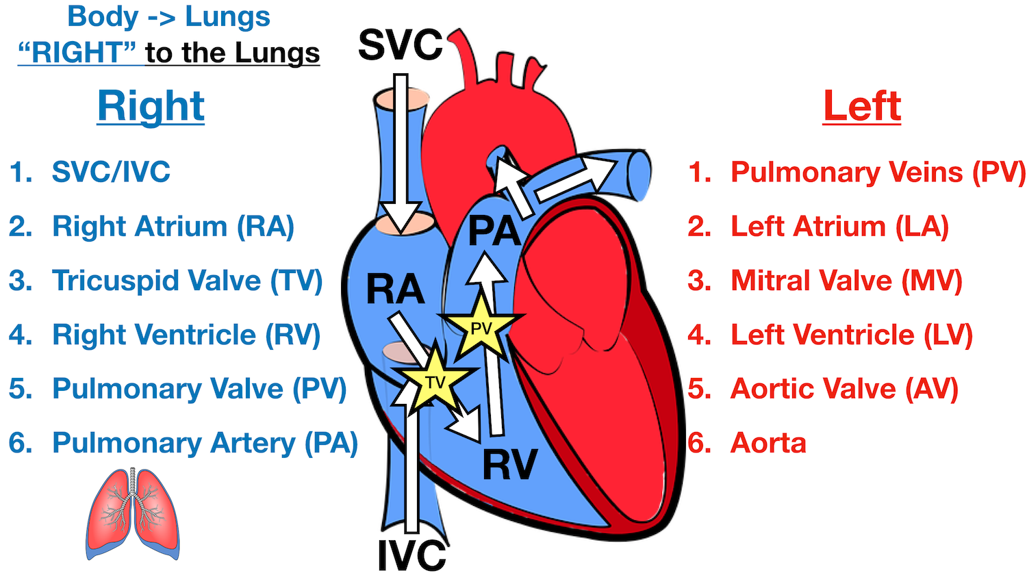
Blood Flow Through the Heart - Registered Nurse RN The un-oxygenated blood (this is blood that has been "used up" by your body and needs to be resupplied with oxygen) enters the heart through the SUPERIOR AND INFERIOR VENA CAVA. 2. Blood enters into the RIGHT ATRIUM. 3. Then it is squeezed through the TRICUSPID VALVE. 4. Blood then enters into the RIGHT VENTRICLE. 5.
Heart Diagram Flow Teaching Resources | Teachers Pay Teachers Cardiovascular System: Heart Diagram to Color by Lori Maldonado 80 $2.00 PDF This diagram shows the way blood flows through the heart. The areas of the heart with MORE oxygen are labeled with an "R". Students will color these areas RED. The areas of the heart with LESS oxygen are labeled with a "B". Students will color these areas BLUE.
Box Diagram, Labels of Heart, and Blood Flow through Heart About Press Copyright Contact us Creators Advertise Developers Terms Privacy Policy & Safety How YouTube works Test new features Press Copyright Contact us Creators ...
Heart Diagrams for Labeling and Coloring, With Reference Chart and Summary (Not for black and white photocopying). - One black and white heart diagram with lines for students to fill in labels, and arrows showing blood flow - One black and white heart diagram with no lines or labels, but arrows included, so you can customize what labels the diagram will include
How the Heart Works How Blood Flows through the Heart Arteries take blood away from your heart. Your heart valves help control the direction the blood flows. Heart valves Heart valves control the flow of blood so that it moves in the right direction. The valves prevent blood from flowing backward. The heart has four valves. The tricuspid valve separates the right atrium and right ventricle.
Human Heart Diagram Labeled | Science Trends Let's examine the anatomy of the heart along with some diagrams that show how the heart operates. Anatomy Of The Heart The human heart usually weighs somewhere between 10 to 12 ounces in men and between 8 to 10 ounces in women, and in terms of size is roughly the size of the fist.















Post a Comment for "38 heart diagram with labels and blood flow"