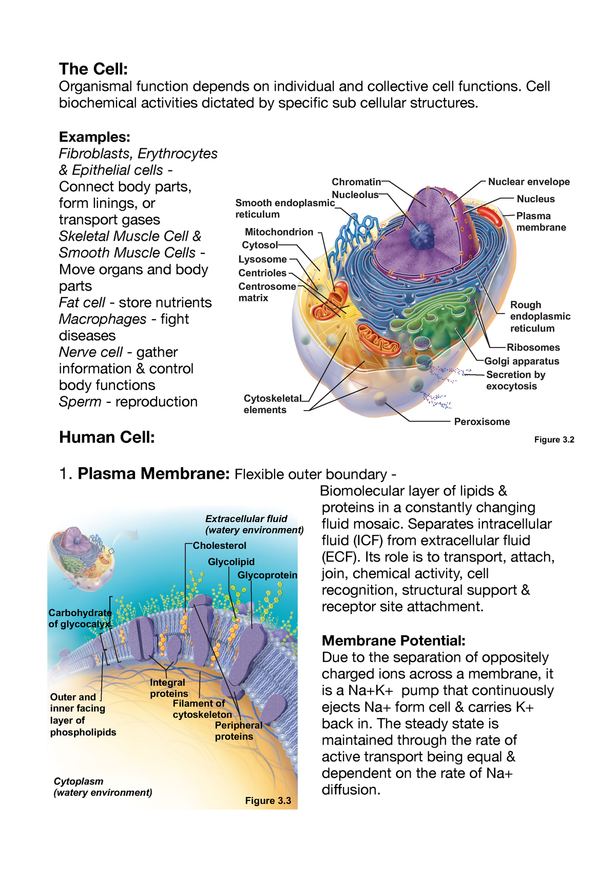
45 cell membrane diagram with labels
DP Biology: Cells: Activities for Learning - ThinkIB Membrane Structure and Transport - planning sheet This simple sheet sets out the learning objectives, essential questions and some ideas for assessment for the following activities. Membrane Structure and function Time: 1h Students will learn how to draw and label a diagram of the fluid mosaic model of membranes. Conformational change of Syntaxin-3b in regulating SNARE complex ... Syntaxin-3b is enriched in the presynaptic plasma membrane of synaptic ribbon synapses and has been shown to be an essential component for synaptic vesicle exocytosis in the retinal bipolar cells ...
Messenger RNA (mRNA) - Genome.gov 00:00. 00:39. Messenger RNA (abbreviated mRNA) is a type of single-stranded RNA involved in protein synthesis. mRNA is made from a DNA template during the process of transcription. The role of mRNA is to carry protein information from the DNA in a cell's nucleus to the cell's cytoplasm (watery interior), where the protein-making machinery ...

Cell membrane diagram with labels
Targeted delivery of fat extract by platelet membrane-cloaked ... In Vitro effect of RGD-PLT@PLGA-FE on tube formation and migration of HUVECs. To evaluate the targeting ability of RGD-PLT@PLGA-FE toward inflamed endothelial cells and their effects on tube formation and migration of HUVECs in vitro, HUVECs were treated with LPS (2 µg/ml) to mimic in vivo inflammation and then incubated with 3,3′-dioctadecyloxa carbocyanine perchlorate (DiO) labeled-PLGA ... › cell_cycle_jsInteractive Cell Cycle - CELLS alive INTERPHASE. Gap 0. Gap 1. S Phase. Gap 2. MITOSIS . ^ Cell Cycle Overview Cell Cycle Mitosis > Meiosis > Get the Cell Division PowerPoints en.wikipedia.org › wiki › Nuclear_envelopeNuclear envelope - Wikipedia The outer nuclear membrane is continuous with the endoplasmic reticulum membrane. The nuclear envelope has many nuclear pores that allow materials to move between the cytosol and the nucleus. [4] Intermediate filament proteins called lamins form a structure called the nuclear lamina on the inner aspect of the inner nuclear membrane and give ...
Cell membrane diagram with labels. Biofabricated macrophage and fibroblast membranes synergistically ... In the photographs acquired with a digital camera (Figure 4b), simulation diagrams ... The results indicated that the combination of cell membrane and TGF-β inhibitor promoted the wound healing. ... the tissues were cut into pieces and digested with trypsin and collagenase IV, and the cells were labeled with anti-CD45-APC-Cy7, anti-CD11b-FITC ... InterCellDB: A User‐Defined Database for Inferring Intercellular ... The spatial pattern of target protein pairs was shown in a schematic diagram. Two separate cell-mimic areas represented two independent cells. Each cell area contained four subcellular locations, including extracellular space, cytomembrane, cytoplasm, and cell nucleus. Interaction type and effect were denoted by line color and arrow. Mitosis in Onion Root Tips (Theory) : Cell biology Virtual Lab II ... Interphase is mainly divided into three phases: G1 phase, S phase and G2 phase. S phase is the period of replication. G1 and G2 are the two gap phases during which the cell grows, producing proteins and preparing the cells. These phases also have certain check points and the whole cell cycle is strictly regulated. chemstuff.files.wordpress.com › unit-1-cellsUnit 1 Biology and Disease Cell structure & function Practice ... The diagram shows the structure of the cell-surface membrane of a cell. Name A and B (2 marks) C is a protein with a carbohydrate attached to it. This carbohydrate is formed by joining monosaccharides together. Name the type of reaction that joins monosaccharides together. Some cells lining the bronchi of the lungs secrete large amounts of mucus.
Circulatory System Diagram - New Health Advisor Coronary circuit mainly consists of cardiac veins including anterior cardiac vein, small vein, middle vein and great (large) cardiac vein. There are different types of circulatory system diagrams; some have labels while others don't. The color blue stands for deoxygenated blood while red stands for blood which is oxygenated. How to Neutralise Glyphosate (Roundup) Herbicide Contamination in Soil That's where the figure on the label of the consumer glyphosate product came from. On the other hand, with a worst-case scenario 197-day half-life, it will take 985 days (2.7 years) for the glyphosate to break down to 3% or the original amount, and using the 0.75% remaining figure used earlier, would be 7 half-lives, or 1379 days (3.8 years). Gram Stain Technique (Theory) : Microbiology Virtual Lab I ... The decolorized Gram negative cells can be rendered visible with a suitable counterstain, which is usually positively charged safranin, which stains them pink. Pink colour which adheres to the Gram positive bacteria is masked by the purple of the crystal violet ( Basic fuschin is sometimes used instead of safranin in rare situations). Animal Plant Cell Diagram - b564abrahamwoods.blogspot.com Plant Cell Diagram Animal Cell Diagram Featured in this printable worksheet are the diagrams of the plant and animal cells with parts labeled vividly. A Guide to Understand Plant Cell with Diagram. Students write the name of the cell parts in the boxes. In your INB you will need to make sure your diagram and chart are completed.
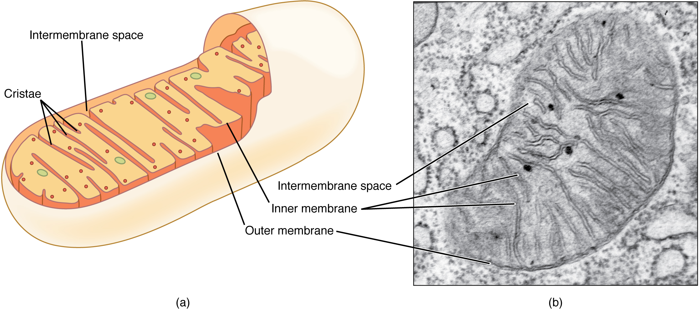
sciencequiz.net › newjcscience › jcbiologyThe Cell - ScienceQuiz.net The diagram shows a plant cell as seen under a microscope. Two of the labels are incorrect. What are they? ... A is the cell membrane and DNA is located inside B.? NMDA receptor - Wikipedia The N-methyl-D-aspartate receptor (also known as the NMDA receptor or NMDAR), is a glutamate receptor and ion channel found in neurons.The NMDA receptor is one of three types of ionotropic glutamate receptors, the other two being AMPA and kainate receptors.Depending on its subunit composition, its ligands are glutamate and glycine (or D-serine).However, the binding of the ligands is typically ... Mitochondria - National Human Genome Research Institute Home Mitochondria are membrane-bound cell organelles (mitochondrion, singular) that generate most of the chemical energy needed to power the cell's biochemical reactions. Chemical energy produced by the mitochondria is stored in a small molecule called adenosine triphosphate (ATP). Mitochondria contain their own small chromosomes. animal cell microscope slide - Veronique Daugherty Two slides demonstrating the cell membrane of an animal. Login or Register 8003345551. Find out the oral epithelial cells with a flat oval. ... As you can see in the above labeled plant cell diagram under light microscope there are 13 parts namely Cell membrane. Avantik - Trusted supplier since 1971 offering high quality consumables. DNA Book ...
Cell Membrane Facts Labeled - Cell Diagram
A Plant Cell Plant Cell is an eukaryotic cell primarily involved in photosynthesis and having its genomic content present in a membrane bound cell organelle ie. Plant cells are a type of eukaryotic cell that are found in organisms of the Plant Kingdom. Plant cells are unique among eukaryotic cells because they are capable of creating their own food.
Flashcards Table on Biology Revision.
What is Rhesus (Rh) typing? - Medscape One gene, RHD, encodes for the D antigen. Individuals with the D antigen present on their red blood cells are labeled as "Rh (D)-positive." Those who do not have the D antigen are labeled as "Rh ...

Cell Membrane Facts Labeled - Cell Diagram
› bitesize › guidesAnimal cells and plant cells - Cells to systems - KS3 ... - BBC Part Function Found in; Cell membrane: Controls the movement of substances into and out of the cell: Plant and animal cells: Cytoplasm: Jelly-like substance, where chemical reactions happen
Label The Following Parts Of The Cell Membrane - img-whammy
quizlet.com › 244659812 › cell-bio-ch-22-flash-cardsCell Bio - Ch. 22 Flashcards | Quizlet The diagram below shows the five main transport proteins that control the distribution of Na+ and K+ ions across the plasma membrane of an axon. Assume that the membrane is at resting potential---the membrane potential of the axon remains constant at about -70 mV.
Biology Websites - Mrs. O' Hehir's Biology Site
Novel Anti-LY6G6D/CD3 T-Cell-Dependent Bispecific Antibody for the ... Crystal structure of LY6G6D-1G4 Fab complex at 2.2 Å resolution suggests Fab binds an epitope located close to the membrane. A, Ribbon diagram of one crystallographic dimer shown from a top view. The complex crystallized in space group P 1 21 1 with two dimers per asymmetric unit; the four complexes within the asymmetric unit superimpose ...

Active and Passive Transport - Difference and Comparison | Diffen
What Do Ribosomes Look Like In A Plant Cell - awhatdok O Cell Membrane - Pink o Cytoplasm - Yellow o Vacuole Light Black o Nucleus - Blue o Mitochondria - Red o Ribosomes - Brown o Endoplasmic Reticulum - Purple o Lisosome Light Green o Golgi Body Orange 2. Endoplasmic reticulum with attached ribosomes is called rough ER. The functions of ribosomes in plant cells are.

Animal Cell by Amy Conley
Cell Organelles Worksheet Answers - dontrabajo.com Together, the absolute breadth of a cell's centralized membranes far exceeds that of its claret membrane. Like the claret membrane, organelle membranes action to accumulate the central "in" and the alfresco "out." This administration permits altered kinds of biochemical reactions to booty abode in altered organelles.
A Detailed Diagram Models Of Cell Membrane Stock Vector Illustration 376416385 : Shutterstock
Diagram of Human Heart and Blood Circulation in It The outermost layer of your heart wall is called the epicardium, which is basically a very thin layer of serous membrane. The membrane provides lubrication and protection to the outer side of your heart, as you can see in heart diagram labeled. Myocardium Right beneath epicardium is another relatively thicker layer called myocardium.
The Cytoplasm and Cellular Organelles · Anatomy and Physiology
Animal And Plant Cell Quiz - ProProfs What is the Cell membrane? A. A thin, flexible barrier around the cell that regulates transport. B. A rigid cover that provides support for the cell. C. The place where light energy, water, and carbon dioxide are used. D. Converts solar energy to chemical energy. 3. What is the primary function of the Cell Wall? A.

Cell membrane with labeled educational structure scheme vector illustration | Cell membrane ...
Dressing blood-contacting devices with platelet membrane ... - cell.com To prepare the PMC, we first stripped the platelet membrane to prepare platelet vesicles (PMVs) with a hydration particle size of about 100 nm ( Figure S1 A) and a ζ potential of −27.1 ± 0.8 mV ( Figure S1 B) as established before. 9 PMVs were then observed by transmission electron microscopy (TEM) ( Figure S1 C).
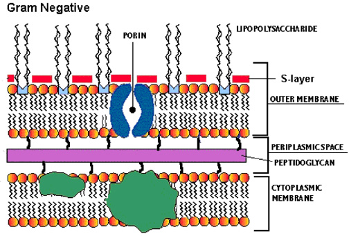
Proteobacteria - microbewiki
en.wikipedia.org › wiki › History_of_cell_membraneHistory of cell membrane theory - Wikipedia Two experiments in 1924 laid the groundwork to fill in this gap. By measuring the capacitance of erythrocyte solutions Fricke determined that the cell membrane was 3.3 nm thick. Although the results of this experiment were accurate, Fricke misinterpreted the data to mean that the cell membrane is a single molecular layer.
Cell Membrane Articles 2018 Structure - Cell Diagram
Transcription factor - Wikipedia In molecular biology, a transcription factor (TF) (or sequence-specific DNA-binding factor) is a protein that controls the rate of transcription of genetic information from DNA to messenger RNA, by binding to a specific DNA sequence. The function of TFs is to regulate—turn on and off—genes in order to make sure that they are expressed in the desired cells at the right time and in the right ...

Cell Membrane Real Life Example Labeled - Cell Diagram
A Plant Cell - c643mitchellwhite.blogspot.com Plant Cell is an eukaryotic cell primarily involved in photosynthesis and having its genomic content present in a membrane bound cell organelle ie. Plant cells are multicellular eukaryotic cells that make up a plant a group of eukaryotes belonging to the Plantae kingdom with the ability to synthesis their own food using water Sunlight and CO2.

Cell Membrane Game Labeled - Cell Diagram
Pick Up Line For Nucleolus - card-stud-software-uckr Parts Of A Cell And Their Functions Cell Membrane Cell Cell Parts Psychology Pick Up Lines.. Answer 1 of 2. Youre so hot you denature my proteins. I must be a snowflake because Ive fallen for. My creations are positioned on the RER and help with protein synthesis and movement throughout. Some interesting pick up lines were tossed around during ...
PPT - Lesson Objectives—Cell Cycle PowerPoint Presentation, free download - ID:9184287
drosophila life cycle diagram - Mireille Hsu Cycle 14 in which the Drosophila embryo forms cells ie after 13 divisions is asynchronous. An XY pair and three autosomes labeled 2 3 and 4. Drosophila is a model organism particularly used in developmental biology because it is a holometabolous insect with major morphological differences occurring between larvae and adult animal metamorphosis ...
Red Blood Cell (RBC) – Part 4 – Erythropoiesis, Red blood cell Count and Counting Procedure ...
Pollen wall and tapetal development in Cymbalaria muralis: the role of ... The tapetal cells appear normal, with exception of columella-like surface pattern as a row of straight short cylindrical micelles (Fig. 4a-d). Somewhat later in development, when the cytoplasm of these tapetal cells with abnormal surface pattern degenerate, the columella-like envelope persists (Fig. 4e, arrows).

Confocal laser scanning microscopy of plant cells labeled with various... | Download Scientific ...
Animal & Plant Cells Quiz For 6th Grade - ProProfs Cell membrane C. Cytoplasm D. Chromosomes E. Cellulose 6. True or False: Cell parts called genes determine what traits a living thing will have. A. True B. False 7. Blood cell that helps destroy germs A. Red blood cell B. White blood cell C. Exterminatory cell D. Squamish cell 8. Movement of water through a membrane A. Water surge B.
Post a Comment for "45 cell membrane diagram with labels"