44 images of compound microscope with labels
Parts of a Compound Microscope - Labeled (with diagrams) Image 3: A compound microscope with a corresponding label of the different parts. imagesource: images.slideplayer.com The optical components of a compound microscope. Image 4: Eyepieces and objective lenses with corresponding magnifications. image source: Eyepiece/ocular What is a Compound Microscope? - New York Microscope Company A compound microscope is an instrument that is used to view magnified images of small specimens on a glass slide. It can achieve higher levels of magnification than stereo or other low power microscopes and reduce chromatic aberration. It achieves this through the use of two or more lenses in the objective and the eyepiece.
Place the labels on the image of a compound light | Chegg.com Expert Answer. Transcribed image text: Place the labels on the image of a compound light microscope. Group 1 labels identify the parts; group 2 labels identify the functions. Coaxial stage controls Stage clip Secures the slide on the stage Move the slide night eft and backwardforward Group 1 Group 2 Group 1 Group 2 Reset Help. Previous question.

Images of compound microscope with labels
Looking at the Structure of Cells in the Microscope For deconvolution, we first obtain a series of (blurred) images, focusing the microscope in turn on a series of focal planes—in effect, a blurred three-dimensional image. The stack of images is then processed by computer to remove as much of the blur as possible. Essentially the computer program uses the microscope's point spread function to determine what the effect of the … Compound Microscope Parts - Labeled Diagram and their Functions - Rs ... Basically, compound microscopes generate magnified images through an aligned pair of the objective lens and the ocular lens. In contrast, "simple microscopes" have only one convex lens and function more like glass magnifiers. [In this figure] Two "antique" microscopes played significant roles in the history of biology. Labeling the Parts of the Microscope | Microscope World Resources Labeling the Parts of the Microscope. This activity has been designed for use in homes and schools. Each microscope layout (both blank and the version with answers) are available as PDF downloads. You can view a more in-depth review of each part of the microscope here.
Images of compound microscope with labels. Compound Microscope Labeled Diagram - Quizlet QUESTION. The total magnification of a specimen being viewed with a 10X ocular lens and a 40X objective lens is. 15 answers. QUESTION. a mosquito beats its wings up and down 600 times per second, which you hear as a very annoying 600 Hz sound. if the air outside is 20 C, how far would a sound wave travel between wing beats. 2 answers. Compound Microscope - Diagram (Parts labelled), Principle and Uses A compound microscope basically consists of optical and structural components. Within these two systems, there are multiple components within them and they are: Image : Labeled Diagram of compound microscope parts See: Labeled Diagram showing differences between compound and simple microscope parts Structural Components en.wikipedia.org › wiki › Electron_microscopeElectron microscope - Wikipedia An electron microscope is a microscope that uses a beam of accelerated electrons as a source of illumination. As the wavelength of an electron can be up to 100,000 times shorter than that of visible light photons , electron microscopes have a higher resolving power than light microscopes and can reveal the structure of smaller objects. Compound microscope Images, Stock Photos & Vectors - Shutterstock 3,117 compound microscope stock photos, vectors, and illustrations are available royalty-free. See compound microscope stock video clips Image type Orientation Sort by Popular Science College and University Biology Insects and Spiders Jobs/Professions microscope laboratory compound eye optical microscope scientist Next of 32
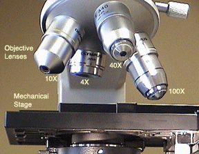
rohrreinigung-notfallservice.de › xawgjpguei › leafLeaf Cell Under Microscope Labeled Jun 19, 2022 · We use the phrase "with the naked eye" to explain that we Robert Hooke was the first cytologist to identify cells under his microscope in 1665. An unknown cell will placed at Station 4 in the back of the classroom. Images were taken on an inverted compound microscope using a 40x DIC objective and digital camera. Compound microscope - their parts and function - Microscopy4kids Compound microscopes have more than one lens to generate high magnification images of flat, thin specimens. 2. Eyepiece (10x) and Objective lenses (4x, 10x, 40x, 100x) are two major optical parts of a microscope. 3. Total magnification power is calculated by multiplying the magnification of the eyepiece and objective lens. 4. rsscience.com › stereo-microscopeParts of Stereo Microscope (Dissecting microscope) - Rs' Science The difference between Compound and Stereo (Dissecting) Microscope. Unlike a compound microscope that can only see a very thin specimen, stereo microscopes can be used for viewing almost anything you can fit under them. However, stereo microscopes offer lower magnification, typically 5x-50x, comparing to compound microscopes. 10 Best Compound Microscopes (Summer 2022) - The Complete Guide Compound microscope is a type of optical microscope that is used for obtaining a high-resolution image. There are more than two lenses in a compound microscope. Learn about the working principle, parts and uses of a compound microscope along with a labeled diagram here.
300+ Free Microscope & Laboratory Images - Pixabay Find your perfect microscope image. Free pictures to download and use in your next project. 189 37. analysis biochemistry. 335 71. analysis biochemistry. 334 96. microscope slide. 725 186. › cemf › whatisemWhat is Electron Microscopy? - UMASS Medical School Conventional scanning electron microscopy depends on the emission of secondary electrons from the surface of a specimen. Because of its great depth of focus, a scanning electron microscope is the EM analog of a stereo light microscope. It provides detailed images of the surfaces of cells and whole organisms that are not possible by TEM. Microscope Images at Various Magnifications | Microscope World Resources The different images below were taken with two different types of microscopes. The images of Paulownia wood, hair, and frog's blood were captured with a high power compound microscope using a Nikon camera adapter. The compound microscope typically has three or four magnifications - 40x, 100x, 400x, and sometimes 1000x. Solved Label the image of a compound light microscope using - Chegg Experts are tested by Chegg as specialists in their subject area. We review their content and use your feedback to keep the quality high. Transcribed image text: Label the image of a compound light microscope using the terms provided.
Microscope World Blog: Biological Microscope Magnifications
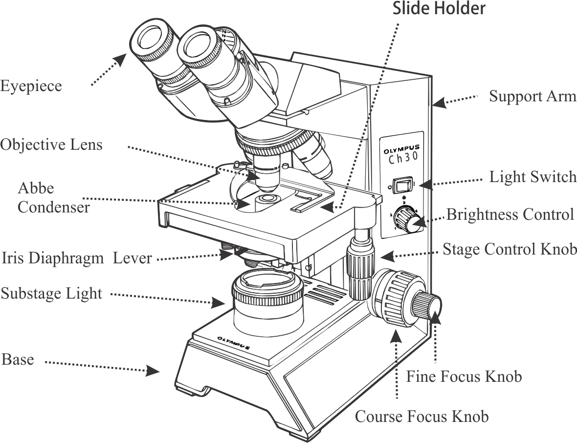
16 Parts of a Compound Microscope: Diagrams and Video In compound microscopes with two eye pieces there are prisms contained in the body that will also split the beam of light to enable you to view the image through both eye pieces. 2. Arm The arm of the microscope is another structural piece. The arm connects the base of the microscope to the head/body of the microscope.

How to Use a Compound Microscope: 11 Steps (with Pictures)
Microscope Components - Science Quiz - GeoGuessr Microscope Components - Science Quiz: The most common type of modern microscope is called a compound microscope. They have two systems of lenses, one is the eyepiece and the other is comprised of one or more objective lenses. This type of microscope has become so advanced that some are capable of magnifying up to 1000 times! Microscopes are used in …

compound microscope 8th standard biology - YouTube
› seterra › en-anMicroscope Components - Science Quiz - GeoGuessr Microscope Components - Science Quiz: The most common type of modern microscope is called a compound microscope. They have two systems of lenses, one is the eyepiece and the other is comprised of one or more objective lenses. This type of microscope has become so advanced that some are capable of magnifying up to 1000 times! Microscopes are used in almost all types of scientific research, and ...

microscope - Kids | Britannica Kids | Homework Help
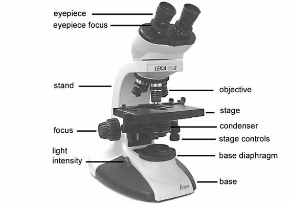
Microscope Types (with labeled diagrams) and Functions A compound microscope: Is used to view samples that are not visible to the naked eye Uses two types of lenses - Objective and ocular lenses Has a higher level of magnification - Typically up to 2000x Is used in hospitals and forensic labs by scientists, biologists and researchers to study micro organisms Compound microscope labeled diagram
The Cell
What is a Compound Microscope? - Microscope Clarity A compound microscope utilizes a system of compounding lenses that enables the microscope to produce highly magnified images. Some of the lenses involved in this compound lens structure are the condenser lens, objective lens (which are themselves made up of several lenses), and the eyepiece lens. Compound microscopes can produce images magnified anywhere from 40x - 2,500x.

Instructions - Microbiology Action - 78 Steps Health
What is Electron Microscopy? - UMASS Medical School Because of its great depth of focus, a scanning electron microscope is the EM analog of a stereo light microscope. It provides detailed images of the surfaces of cells and whole organisms that are not possible by TEM. It can also be used for particle counting and size determination, and for process control. It is termed a scanning electron microscope because the image is formed by …
8 Best Images of Lens Diagram Worksheet - Microscope with Labeled Parts, Label Eye Parts ...
Compound Microscope: Definition, Diagram, Parts, Uses, Working ... - BYJUS A microscope with a high resolution and uses two sets of lenses providing a 2-dimensional image of the sample. The term compound refers to the usage of more than one lens in the microscope. Also, the compound microscope is one of the types of optical microscopes. The other type of optical microscope is a simple microscope.

Microscope World Blog: Radiolaria under the Microscope
Parts of Stereo Microscope (Dissecting microscope) – labeled … Compared to a compound microscope where the objectives attached to the nosepiece can be seen and identified individually (based on color bands and their respective labels), the objectives of a dissecting microscope are located in a cylindrical cone and, therefore, are not directly seen. For the stereo microscope that comes with multiple objective lens sets (fixed power style), the …

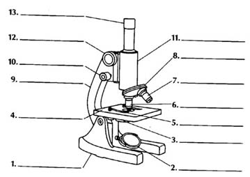
Label diagram of compound microscope - Science - The Fundamental Unit of Life - 12499729 ...
Tirth27/Skin-Cancer-Classification-using-Deep-Learning In the data pre-processing steps, all images are cropped into 768x786 and 512x512 resolution to reduce random noise on the edges of the image. The data cleaning and pre-processing step are performed on all the dataset obtained from the 2020, 2019 and 2018 competition. Also, the image labels are reconciled and combined into a single training CSV ...

labeled microscope for kids - Google Search | {School} - Science | Biology for kids, Teaching ...
Compound Microscope with labels Stock Vector | Adobe Stock Get 10 free Adobe Stock images. Start now Compound Microscope with labels
Labeled Compound Microscope - ClipArt Best
Food Calorimetry: How to Measure Calories in Food We have the compound microscope you are looking for! Digital Microscopes. Digital microscopes are great for large classroom computer combined instruction. Students can take images, videos, and more. Stereomicroscopes. Stereomicroscopes show 3D images vs. flat images and are easier to focus and use. They are great for first tme student use. Physical & …

Mrs. Conner's Science Place: Microscope Parts
Compound Microscope - Types, Parts, Diagram, Functions and Uses Image 5: The arm of the compound microscope. Picture Source: zfic.org Arm - it supports the head of the microscope and attach it to the base. Image 6: The revolving nosepiece. Picture Source: img-aws.ehowcdn.com Nosepiece - It holds the objective lens and attaches them to the head of the microscope.
Compound Microscope Unlabeled - Micropedia
Parts of a Compound Microscope and Their Functions The main parts of compound microscope are the condenser lens, the objective lens, and the eyepiece lens, and these instruments are referred to as compound microscopes. Each of these components is made up of microscope lens combinations that are required to produce magnified images with minimal artefacts and aberrations. Structure of Microscope
Introduction to the light microscope Flashcards | Easy Notecards
931 Compound Microscope Photos - Free & Royalty-Free Stock Photos from ... Browse 931 professional compound microscope stock photos available royalty-free. Microscope in blue science medical technology laboratory background. Compound microscope in blue science medical technology laboratory background. Typical animal cell Center 400x. The typical animal cell can be seen here. Cell membrane, nucleus, and cytoplasm are ...

Labeled Compound Microscope - ClipArt Best
Cell Types Gizmo Worksheet - StuDocu Gizmo Warm-up In the Cell Types Gizmo, you will use a light microscope to compare and contrast different samples. On the LANDSCAPE tab, click on the Elodea leaf. (Turn on Show all samples if you can’t find it.) Switch to the MICROSCOPE tab to observe the sample as it would appear under the microscope. By default, this microscope is using 40x ...
Post a Comment for "44 images of compound microscope with labels"